Journal of Sol-Gel Science and Technology, 2017, vol 83, 2, pp. 355-364
DOI:10.1007/s10971-017-4428-6
Abstract
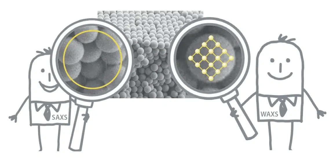
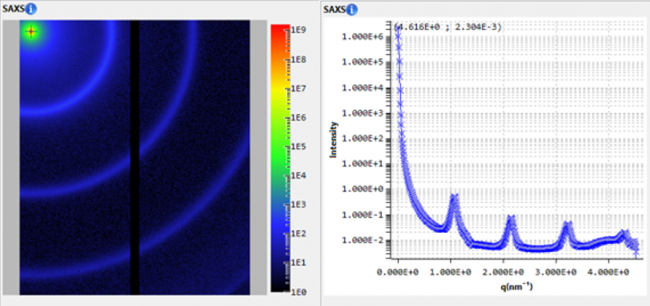
Mesoporous silica nanoparticles were prepared in aqueous/organic phase using cetyltrimethylammonium bromide and polystyrene as organic templates. The morphology and crystalline phase of the products were characterized by scanning electron microcopy, transmission electron microscopy, X-ray diffraction, small angle X-ray scattering, and N2 adsorption/desorption isotherm analysis. The octane/water ratio influenced the pore size distribution, the morphology and size of the nanospheres obtained. Transmission electron microscopy revealed that mesoporous silica nanoparticles with “blackberry-like structure” (MSN3, MSN4, MSN5, and MSN6 samples) were obtained using octane/water ratios in the range 0.007–0.35. They present small (in the range 5–6 nm) and large (in the range 28–34 nm) mesopores. Large mesopores were mainly generated by polystyrene, and their volume contribution was clearly higher than in the MSN1 and MSN2 samples. The structure and morphology of mesoporous silica nanoparticles solids impregnated with tungstophosphoric acid were similar to those of the mesoporous silica nanospheres used as support. In addition, the characterization of all the solids impregnated with tungstophosphoric acid by Fourier transform infrared and 31P nuclear magnetic resonance indicated the presence of undegraded [PW12O40]3− and [H3−X PW12O40](3−X)− species interacting electrostatically with the ≡Si–OH2 + groups, and by potentiometric titration the solids presented very strong acid sites. In summary, they are good candidates to be used in reactions catalyzed by acids, especially to obtain quinoxaline derivatives.Graphical Abstract Open image in new window


