7-12 July, 2024
Xenocs is pleased to attend the American Crystallographic Association’s 74th Annual Meeting from July 7-12 in Denver, Colorado, USA. Meet us at booth 511 in the exhibitor hall.

Xenocs is pleased to attend the American Crystallographic Association’s 74th Annual Meeting from July 7-12 in Denver, Colorado, USA. Meet us at booth 511 in the exhibitor hall.
Based on worldwide patents and more than 14 years of Research and Development, Xenocs is the only company in the world proposing single reflection multilayer optics. These optics offer typically 50% more efficiency than standard multiple reflection optics, also called Montel optics.
Based on its proprietary replication technology, Xenocs is able to offer innovative optical designs with unmatched performance, which up to now have been impossible to achieve with other existing manufacturing techniques.
| Mirror type | Figure | Optic efficiency (calculated over mirror length for 70µm source) |
| Standard Montel mirrors |  | Eff = 42% |
| Xenocs FOX 3D mirrors |  | Eff = 62% |
| Model | Beam size at focus (mm) with 60 µm source | Divergence (mrad) | Typical application |
| FOX 3D Cu 14-39 (♦) | 0.19 x 0.19 | 5.4 | Protein crystallography (♦) Powder diffraction |
| FOX 3D Cu 12-53 | 0.3 x 0.3 | 3 | Protein crystallography (Long Unit Cells) Powder diffraction |
| FOX 3D Mo 10-31 | 0.13 x 0.13 | 4 | Small molecule crystallography High pressure diffraction |
| FOX 3D Ag 10-30 | 0.13 x 0.13 | 3 | Small molecule crystallography High pressure diffraction |
| FOX 3D Cr 8-30 | 0.20 x 0.20 | 6 | Protein crystallography (S-SAD) Stress analysis |
| FOX 1D Cu 12-53 | 0.3 | 3 | XRD, Powder Diffraction |
| Model | Beam size at focus (mm) with 60 µm source | Divergence (mrad) | Typical application |
| FOX 2D Mo 25-25 (♦) | 0.08 x 0.08 | 5 | Small mollecule crystallography High pressure crystallography |
| FOX 2D Cu 25-25 (♦) | 0.08 x 0.08 | 5.3 | Protein crystallography (♦) Micro Diffraction, Reflectometry |
| FOX 3D Cu 21-21 HC (♦) | 0.08 x 0.08 | 70 x 35 | Rapid x-ray Reflectometry (full beam) Scanning x-ray Reflectometry (with slit) Micro-XRF |
| FOX 3D Cu 28-10 | 0.04 x 0.04 | 17 | Stress Analysis, Micro-diffraction, Micro-XRF |
| FOX 3D Cr 30-8 | 0.04 x 0.04 | 17 | Stress Analysis, Micro-diffraction, Micro-XRF |
| Model | Beam size at focus (mm) with 60 µm source | Divergence (mrad) | Typical application |
| FOX 2D Cu 12-INF (♦) | 1.2 x 1.2 | 1 | Small angle x-ray scattering (♦) High resolution x-ray diffraction (♦) Surface scattering (♦) Thin film analysis (reflectometry stress) (♦) Microdiffraction on synchrotron (♦) |
| FOX 2D Mo 25-INF (♦) | 0.85 x 1.3 | 0.5 | Small angle x-ray scattering High resolution x-ray diffraction Surface scattering |
| FOX 2D 12-60 L (♦) | 0.18 x 0.2 | 0.5 x 2 | High resolution diffraction Small sample analysis |
| FOX 1D Cu 12-INF (♦) | 1.2 | 0.8 | High resolution x-ray diffraction, Powder diffraction, Reflectometry, Small angle x-ray scattering |
| Model | Beam size at focus (mm) with 60 µm source | Divergence (mrad) | Typical application |
| FOX 1D Cu 12-53 | 0.3 | 3 | XRD, Powder Diffraction |
| Model | Beam size at focus (mm) with 60 µm source | Divergence (mrad) | Typical application |
| FOX 1D Cu 12-INF (♦) | 1.2 | 0.8 | High resolution x-ray diffraction, Powder diffraction, Reflectometry, Small angle x-ray scattering |
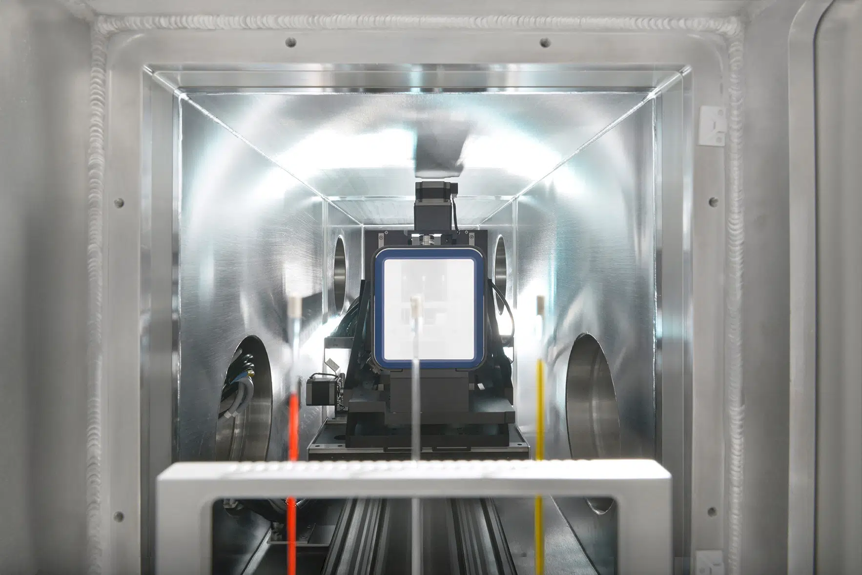
Advanced materials research and characterization could require integration of specific or customized sample environments.
Xeuss 3.0 is perfectly adapted for such new projects thanks to its state of the art beamline concept.
It provides:
In particular, the following key features enable the user to optimize experiments or results:
SAXS acquisition continuously without any beamstop provides High Dynamic Range data.
No parasitic scattering from beamstop edges or detector window allows noise-free scattering at low q.
The direct beam is recorded simultaneously with the sample scattering profile during acquisition for accurate transmitted intensity measurement, used to obtain a precise absolute intensity normalization.
In addition, the direct beam profile thus measured is integrated in the data analysis (XSACT) to improve the accuracy of the results.
As a standard solution, Xeuss 3.0 integrates low maintenance microfocus beam delivery systems to provide very high flux levels previously only possible with high power rotating anode sources.
Advanced experiments such as kinetic studies or shape analysis of diluted samples can now be performed in the lab.
In addition, Xenocs X-ray beam delivery systems coupled with motorized scatterless collimation achieve high resolution (low Δq beam), which is essential for studying large characteristic dimensions or for performing accurate mesophase analysis.
The high useful flux together with the high resolution capability are achieved through the combination of:
To ensure the best quality of X-ray scattering measurements, including on diluted or low contrast samples, Xeuss 3.0 combines high flux with low noise technology, which is the fruit of more than 10 years of development.
Key integrated features are:
For applications requiring very fast kinetics Xeuss 3.0 can also be delivered with a MetalJet source.
The Q-Xoom features full range computer-controlled detector travel to automatically adjust the sample-to-detector distance offering maximum flexibility of measurement for experiment optimization.
The virtual detector increases the measuring capabilities offering surfaces of collection larger than 200 mm2 x 200 mm2 at any sample-to-detector distance independently of detector size with automatic data reconstruction.
The large surface of detection is beneficial to optimize the azimuthal coverage in case of anisotropic samples or to benefit from high resolution measurement settings by characterizing smaller length scales (larger wave vectors) at longer sample to detector distance by a detector offset.
Advanced nanomaterials development and design require characterization over a large range of length scales. The Xeuss 3.0 offers such measuring capability over up to 5 orders in magnitude in q (wave vector) through entirely motorized change of configurations. Any trained user can thus operate the system remotely over its complete measurement range for a given sample or batch of samples.
Automatic change of measuring settings include:
– Q-Xoom change of measurement resolution through motorized translation of detector
– Sequential SAXS /USAXS measurement with Bonse-Hart USAXS module
– Change of energy radiation, up to 3 energies
– Movable WAXS detector for out of equilibrium in situ SWAXS studies
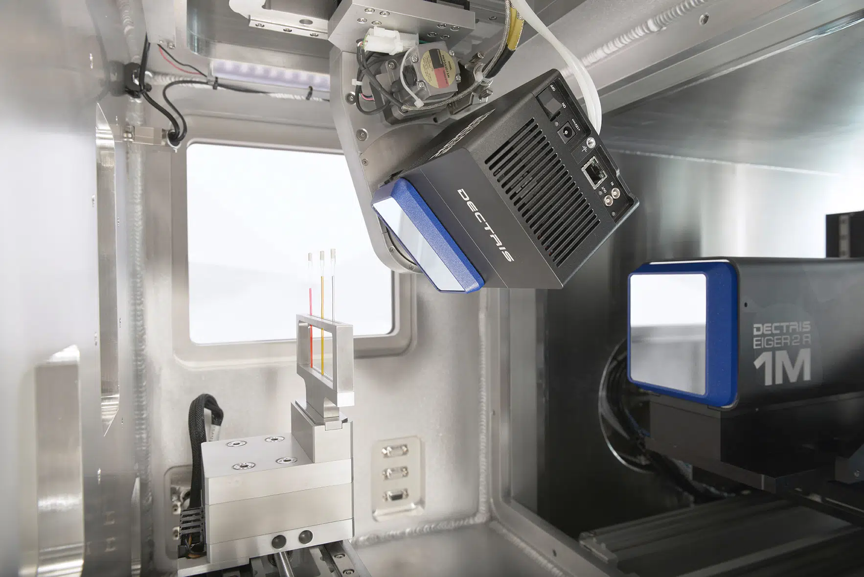
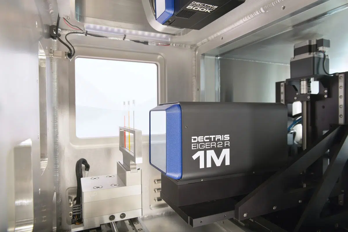
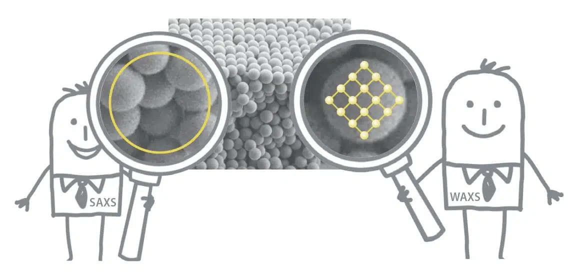
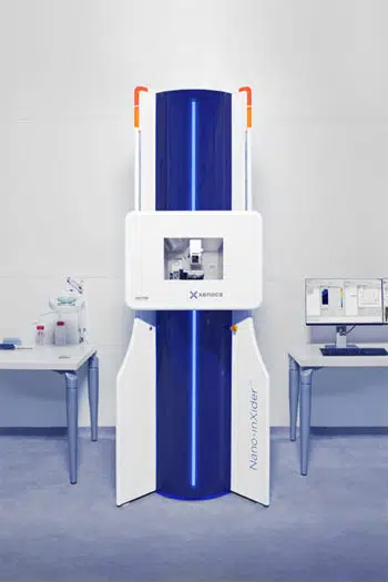
The Nano-inXider features a smart dual detector design to detect atomic scale information and nanostructure simultaneously within one exposure. Long sample-to-detector distance for measuring large characteristic dimensions is achieved in SAXS through a vertical design with a small footprint.
Such configuration provides unique benefits:
With the Nano-inXider, the sample and the detectors are fixed. The optional WAXS detector extends the scattering range from the SAXS detector seamlessly throught 2Ɵ=60°.
Such a unique vertical design provides unique benefits:
For anisotropic samples like fibers or oriented films, the Nano-inXider is equipped with a sample rotation stage that acquires the anisotropic scattering. The automatic acquisition involves the following steps:
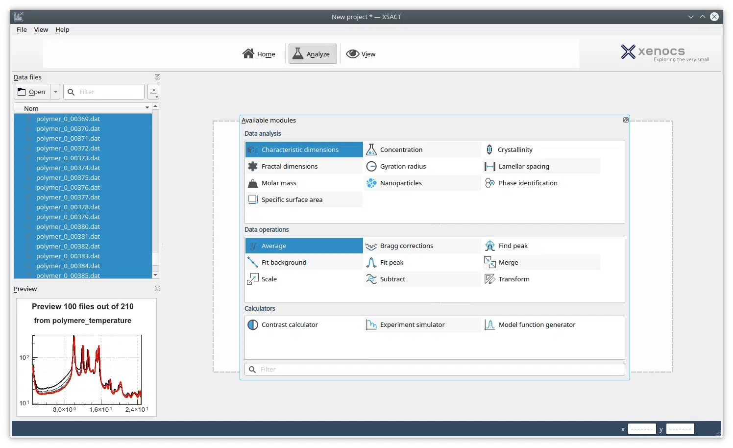
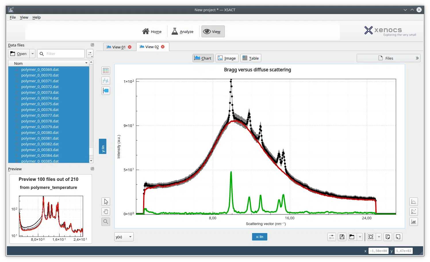
The SAXS community relies on multiple algorithms with custom models and many different applications. XSACT offers a unified, versatile and complete data processing solution to get things done as fast as possible.
The low cost of ownership of the Nano-inXider mainly comes from its compactness and low cost of operation
The Nano-inXider acquires high signal to noise data by measuring the intense direct beam transmitted through the sample together with low intensity signal scattered from the sample.
Direct beam measurement, achieved with innovative beamstop-less data acquisition, enables automatic data treatment and display in absolute intensity with a very high accuracy.
Simultaneous low intensity signals detection is rendered possible by implementation of Clean Beam Technology.
The high dynamic range of intensity collection directly impacts data quality by:
– Enabling the detection of low intensity scattered signal from weakly scattering samples
– Accessing absolute quantity parameters such as particles number, molar mass, concentration, specific surface
– Detecting large characteristic dimensions without need of user data treatment
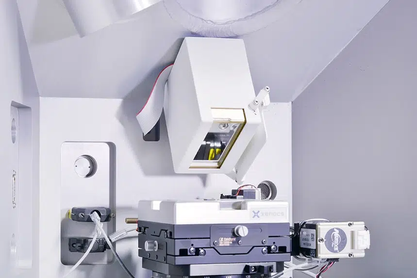
The Nano-inXider integrates 15 years of R&D in the field of advanced components and instrument design to achieve an optimized balance of high X-ray flux on sample together with low parasitic scattering generated by the instrument.
Clean Beam Technology embeds the following key components and features:
The Nano-inXider measures SAXS without any beamstop and simultaneously collects the direct beam transmitted through the sample together with the scattered signal down to very low intensity levels.
Such high dynamic range data collection is rendered possible by innovative beamstop-less data acquisition.
The direct beam is recorded all along the acquisition, which leads to an accurate transmitted measurement used to obtain a precise absolute intensity normalization.
In addition, the measured direct beam profile (resolution function) is integrated in the data analysis to improve results quality.
Xenocs beamstop-less data acquisition integrates automatic qmin determination depending on the sample and benefits from no parasitic scattering from beamstop edges for high data quality at low q.
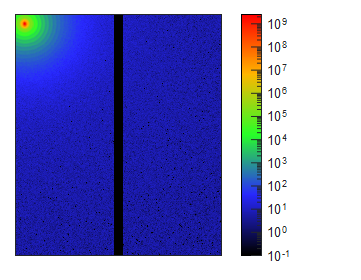
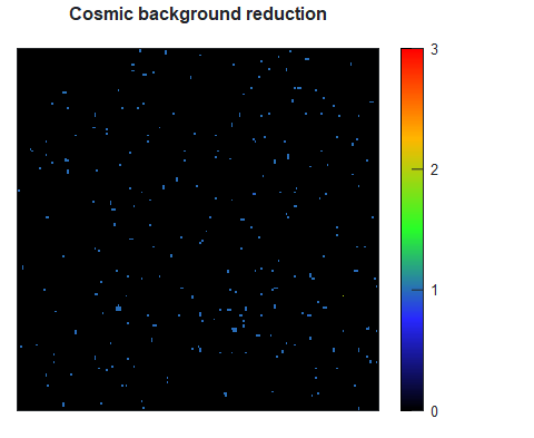
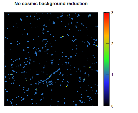
Automatic cosmic background removal reduces the impact of ambient parasitic scattering.
For the best understanding of output data, a scientist must have the tools to visualize, exchange and compare numerous graphical representations.
By combining data processing with advanced visualization capabilities, XSACT speeds up your workflow from acquisition to publication.
With the BioXolver you get:
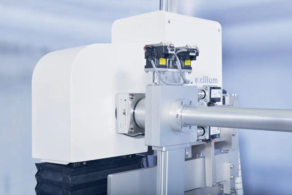
Due to weak scattering from biological samples, high quality SAXS measurements require an instrument optimized for maximum X-ray intensity and lowest possible background noise. The BioXolver offers a unique combination of both capability in particular with:
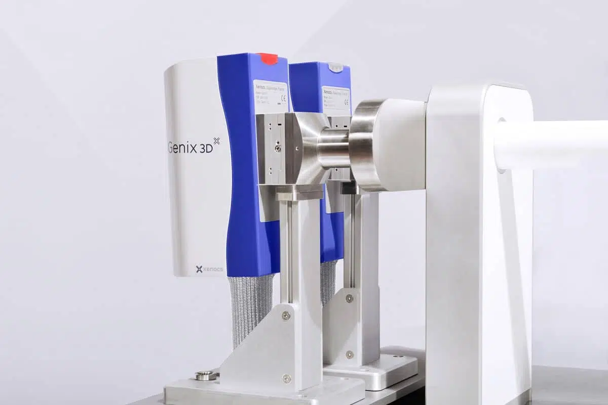
The BioXolver can be installed with different source options.
It can integrate a MetalJet source to obtain the highest X-ray beam intensity possible in a laboratory instrument.
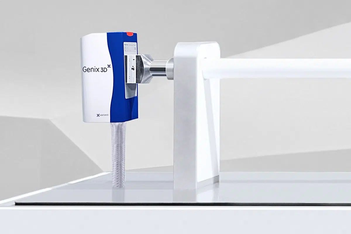
It can also be installed with the Genix3D, a bright microfocus low maintenance source with intensity levels only previously accessible with high power rotating anode sources. Such performance is achieved by combining the Genix3D long lasting source with new patented FOX 3D multilayer optics having a very high solid angle.
The BioXolver comes with windowless hybrid photon counting (HPC) detectors which allows for beamstopless measurements.
By operating without beamstop the direct beam is recorded all along the acquisition, which leads to an accurate transmission measurement used to obtain a precise absolute intensity normalization.
Moreover, operating without beamstop reduces the lowest scattering angle achievable as it is no more limited by beamstop size but is sample dependent.
The BioXolver uses state of the art collimation slits that greatly reduce noise and provide a clean beam.
High quality detection is achieved with hybrid photon counting (HPC) detectors integrated inside vacuum with no interfering window in front of detector sensor (windowless detector). This ensures that weakly scattering samples can also be measured at low concentrations.
Overall measuring background level of the instrument is further improved by proprietary automatic cosmic background removal algorithms suppressing high energy cosmics hitting the detector.
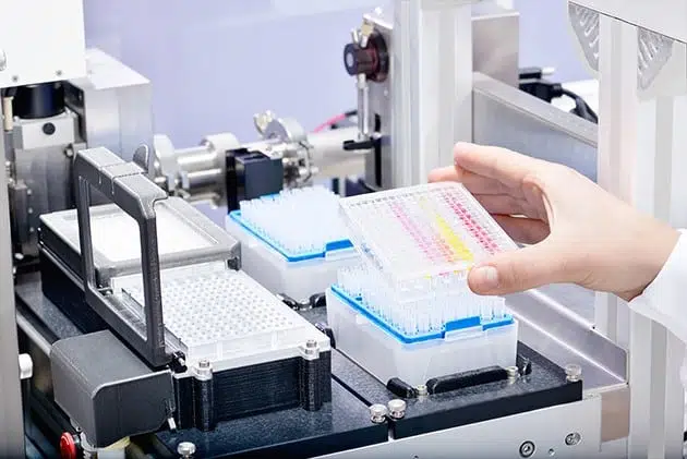
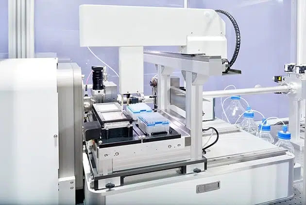
With the BioXolver standard well plates can be loaded & measured automatically using in-line pipetting robot & automated software suite for high user benefits.
The BioXolver includes powerful data reduction BioXTAS RAW widely used by the BioSAXS community.
It enables standard data operation and data analysis (Rg, molecular weight, etc.), together with advanced analysis using ATSAS plugins.
For high throughput analysis, the automated analysis function analyzes and compiles the results for you.
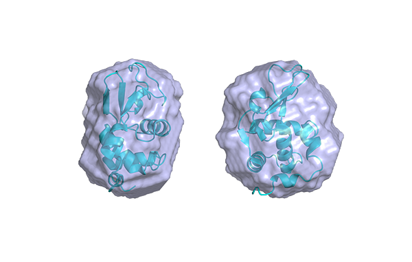
Sample size separation using size-exclusion chromatography in combination with the BioXolver can not only be used to ensure clean monodisperse samples, but also facilitates research on structurally heterogeneous & aggregation-prone multi-domain proteins as well as heterogeneous protein complexes in dynamic equilibria.
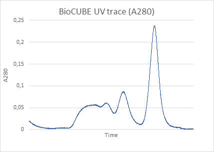
The BioXolver will work with any HPLC system, simply position the HPLC next to the instrument and hook up the column to the BioCUBE flow cell. The in-line UV/Vis ensures accurate determination of the sample concentration exactly at the SAXS exposure position. The included analysis software automatically reduces and provides an easy overview of your entire SEC-SAXS run and makes further analysis easy.
In situ UV measurement directly in the measurement cell provides concentration information of the sample before and after X-ray exposure or during a SEC-SAXS experiment.
This is especially important in this case as the sample is diluted in the transport path from the (HPLC) column exit to the x-ray exposure position.
通过BioXolver,您可以得到:
由于生物样品的微弱散射,高质量的SAXS测量需要一台仪器来达到最大X射线强度和尽可能低的背景噪音。通过以下技术的组合,BioXolver具备了这两种独特的功能:
BioXolver可以配备不同的光源。
可以结合MetalJet光源,在实验室仪器中获得尽可能高的X射线光束强度。
也可以安装Genix3D,一种低维护成本的高亮度微聚焦光源,其强度以前只有高功率阳极转靶才能达到。这种性能通过结合Genix3D耐用光源与具有超高立体角、新的专利FOX 3D多层膜聚焦镜实现。
BioXolver配备了无窗口膜混合光子(HPC)探测器,能够实现无beamstop测试。
在无beamstop情况下,整个测试过程中一直记录直通光,通过准确的透射测试来得到准确的绝对强度校正。
此外,由于不受beamstop尺寸限制,而只与样品有关,无beamstp测试可得到更小的最小散射角度。
BioXolver采用了最先进的准直狭缝,极大地降低了噪音,从而得到了纯净光束。
通过在真空中利用混合光子计数(HPC)探测器,且探测器传感器(无窗口膜探测器)前面无干扰,实现了高质量的检测。这可确保弱散射样品也可以在低浓度下测量。
采用专用的宇宙背景自动去除法,抑制高能宇宙射线,进一步降低了仪器整体的背景噪音。
BioXolver结合了尺寸排阻色谱法来进行区别样品大小,不仅可以保证样品完全单分散,还能促进对结构异质且易于聚集的多域蛋白以及动态平衡中异质蛋白复合物的研究。
BioXolver适用于任何HPLC系统,只需将HPLC置于仪器旁边,并将色谱柱连接到BioCUBE流动池。在线UV/Vis能够确保在SAXS曝光位置精确测量样品浓度。自带的分析软件可自动简化概述整个SEC-SAXS运行过程,这使得进一步分析更加容易。
由于原位UV测试直接在测试池进行,在X射线曝光之前和之后或在SEC-SAXS测试中都能得到样品的浓度信息。
因为样品在从(HPLC)柱出口到X射线曝光位置的传输过程中会被稀释,所以在这种情况下,这一点尤为重要。
通过内置移液机械臂,毛细管流动池BioCUBE以及机械视觉的结合对样品进行监测,确保了少量样品的高精度定位。
在不同环境或浓度下样品总消耗低,对生物结构研究非常有益。
无管操作确保样品消耗量低至5 μl。这在同类仪器中是独一无二的。
每个样品首先由无管移液机械臂自动注入到BioCUBE测量池中,然后定位到光束中进行测量,并实时对其位置进行连续控制。
样品在孔盘中储存期间和在BioCUBE测试期间都始终保持在选定的温度下。
您还可以选择具有集成UV/Vis功能的BioCUBE。
利用机械视觉技术,BioXolver能够随时对样品进行数字化监控。
样品装载后就会被准确地定位在X射线光束中,同时持续反馈结果给自动控制软件来保持光束中的样品量不变。
SAXS用户群倾向于具有自定义模型和多种不同应用程序的算法。XSACT提供的统一、全面且完整的数据处理解决方案可尽快完成任务。
Nano-inXider独特的智能化双探测器设计,可以在一次曝光内同时检测到原子尺度信息和纳米结构。设备占地面积小的垂直设计,样品到探测器距离长,可以测量大的特征尺寸。
这些配置可以提供以下独特的优势:
Nano-inXider的样品和探测器都是固定的。可选的WAXS探测器配置扩大了散射范围,可以无缝衔接SAXS探测器,得到的数据达到2θ=60°。
这独特的垂直设计有以下优势:
对于纤维或取向薄膜等各向异性样品,Nano-inXider配备一个样品旋转台可以获得各向异性散射信号。自动采集包括以下步骤:
Nano-inXider可以通过测量透过样品的强直通光和样品的低散射强度信号来获得高信噪比数据。
通过先进的无beamstop数据采集进行直通光测量,自动处理数据并获得准确度高的绝对强度。
纯净光技术可以实现同时检测低强度信号和高强度信号。
强度采集的高动态范围直接影响数据质量:
– 能够检测弱散射样品的低强度散射信号
– 定量获得粒子数、摩尔质量、浓度、比表面积等参数
– 探测大特征尺寸,无需用户进行数据处理
Nano-inXider通过在先进组件和仪器设计上长达15年的研发,达到了在样品上高X射线通量与仪器产生的低寄生散射的最佳平衡。
纯净光技术具有以下主要组件和特点:
Nano-inXider无需任何beamstop就可以进行SAXS测量,还可以同时采集透过样品的直通光和非常低强度散射信号。
通过先进的无beamstop数据采集可实现高动态范围的数据收集。
在整个的采集过程中记录直通光,使透射测试更准确,从而使绝对散射强度校正更准确。
另外,被测量的直通光(分辨率函数)会被整合到数据分析中来提高测量结果的质量。
Xenocs无beamstop数据采集实现了取决于样品的自动qmin测量,并且消除了beamstop边缘的寄生散射,从而在低q区域获得高质量数据。
自动去除宇宙背景可以减少环境寄生散射的影响。
先进纳米材料的发展和设计需要在超大纳米尺度范围上进行表征。Xeuss 3.0具备这样的测试能力,通过完全自动改变配置,采集的散射矢量q可跨越5个数量级。因此,对于给定的单个或批量样品,任何经过培训的用户都可以在完整的测量范围内远程操作系统。
可自动更改的测量设置包括:
– Q-Xoom通过探测器沿光路的自动平移来改变测量分辨率
-使用Bonse Hart USAXS模块可以实现连续的SAXS/USAXS测量
– X光源能量可选,提供3个可选光源
– 用于原位SWAXS研究的可移动WAXS探测器
尤其,以下关键特点使用户得以优化最佳的实验和结果:
无beamstop连续采集高动态范围SAXS数据。
杜绝来自Beamstop边缘或探测器窗口膜的寄生散射,使低q处的散射信号不受干扰。。
在采集过程中同时记录直通光和样品散射信号,得到精确的透射强度,用于获取精确的绝对强度校正。
另外,采集的直通光信号将整合到数据分析软件(XSACT),提高解析结果的准确性。
作为一个标准的解决方案,Xeuss 3.0集成低维护成本的高性能微聚焦光源,亮度达到了之前高功率旋转阳极靶光源才能实现的水平。
现在实验室中就可进行诸如动力学研究或稀释样品的纳米粒子形状分析的高级实验。
另外,通过结合Xenocs公司的X射线光束和自动无散射准直系统实现高分辨率(光束Δq低),这对研究大特征尺寸的样品或进行准确中间相的分析都非常重要。
通过以下组合实现高光通量和高分辨率:
为了保证X射线散射的最佳测试质量,包括稀释的或对比度低的样品,Xeuss 3.0将10多年研发的高光通量和低噪音技术相结合。
主要集成功能包括:
配备MetalJet光源,用于非常快的动力学研究。
Q-Xoom采用电脑控制探测器全程移动,自动调节样品到探测器距离,提供最大的测试灵活性来优化实验。
虚拟探测器增强测试能力:在任意的样品到探测器距离下,结合图谱重构技术,任何规格的探测器的采集面积都将大于200 mm2 x 200 mm2。
超大探测面积有利于优化各向异性样品信号的方位角覆盖,还有利于高分辨率测试,例如在距离样品较远处用偏置的探测器采集到更大的散射矢量,以表征较小的尺度。
先进材料的研究和表征需要集成特定的或定制的样品环境。
由于其先进的线站理念,Xeuss 3.0非常适用于这种新项目。
包括:
Xenocs基于全球专利和超过14年的研发,成为世界上唯一一家提供单反射多层聚焦镜的公司。这些聚焦镜的效率通常比标准的多反射聚焦镜(也称为Montel聚焦镜)高出50%。
基于专有的光反射技术,Xenocs能够提供具有强大性能的创新聚焦镜设计,这是迄今为止其他现有制造技术无法实现的。
| Mirror type | Figure | Optic efficiency (calculated over mirror length for 70µm source) |
| Standard Montel mirrors | | Eff = 42% |
| Xenocs FOX 3D mirrors | | Eff = 62% |
| Model | Beam size at focus (mm) with 60 µm source | Divergence (mrad) | Typical application |
| FOX 3D Cu 14-39 (♦) | 0.19 x 0.19 | 5.4 | Protein crystallography (♦) Powder diffraction |
| FOX 3D Cu 12-53 | 0.3 x 0.3 | 3 | Protein crystallography (Long Unit Cells) Powder diffraction |
| FOX 3D Mo 10-31 | 0.13 x 0.13 | 4 | Small molecule crystallography High pressure diffraction |
| FOX 3D Ag 10-30 | 0.13 x 0.13 | 3 | Small molecule crystallography High pressure diffraction |
| FOX 3D Cr 8-30 | 0.20 x 0.20 | 6 | Protein crystallography (S-SAD) Stress analysis |
| FOX 1D Cu 12-53 | 0.3 | 3 | XRD, Powder Diffraction |
| Model | Beam size at focus (mm) with 60 µm source | Divergence (mrad) | Typical application |
| FOX 2D Mo 25-25 (♦) | 0.08 x 0.08 | 5 | Small mollecule crystallography High pressure crystallography |
| FOX 2D Cu 25-25 (♦) | 0.08 x 0.08 | 5.3 | Protein crystallography (♦) Micro Diffraction, Reflectometry |
| FOX 3D Cu 21-21 HC (♦) | 0.08 x 0.08 | 70 x 35 | Rapid x-ray Reflectometry (full beam) Scanning x-ray Reflectometry (with slit) Micro-XRF |
| FOX 3D Cu 28-10 | 0.04 x 0.04 | 17 | Stress Analysis, Micro-diffraction, Micro-XRF |
| FOX 3D Cr 30-8 | 0.04 x 0.04 | 17 | Stress Analysis, Micro-diffraction, Micro-XRF |
| Model | Beam size at focus (mm) with 60 µm source | Divergence (mrad) | Typical application |
| FOX 2D Cu 12-INF (♦) | 1.2 x 1.2 | 1 | Small angle x-ray scattering (♦) High resolution x-ray diffraction (♦) Surface scattering (♦) Thin film analysis (reflectometry stress) (♦) Microdiffraction on synchrotron (♦) |
| FOX 2D Mo 25-INF (♦) | 0.85 x 1.3 | 0.5 | Small angle x-ray scattering High resolution x-ray diffraction Surface scattering |
| FOX 2D 12-60 L (♦) | 0.18 x 0.2 | 0.5 x 2 | High resolution diffraction Small sample analysis |
| FOX 1D Cu 12-INF (♦) | 1.2 | 0.8 | High resolution x-ray diffraction, Powder diffraction, Reflectometry, Small angle x-ray scattering |
| Model | Beam size at focus (mm) with 60 µm source | Divergence (mrad) | Typical application |
| FOX 1D Cu 12-53 | 0.3 | 3 | XRD, Powder Diffraction |
| Model | Beam size at focus (mm) with 60 µm source | Divergence (mrad) | Typical application |
| FOX 1D Cu 12-INF (♦) | 1.2 | 0.8 | High resolution x-ray diffraction, Powder diffraction, Reflectometry, Small angle x-ray scattering |
Xenocs凭借其在多层涂层、表面成型和先进的测量技术方面的丰富经验,开发了一系列高性能的XRF分析仪聚焦镜。
凭借其独特的复型技术,Xenocs已经开发了新的光束分析仪,提高了探测下限,增大了可检测量。
| Element of interest | Reference |
| O, F, Na, Mg, Al, Si, P, S | XAN-1 |
| Nitrogen | XAN-2 |
| Carbon | XAN-3 |
| Boron | XAN-4 |
| Mg, Al , Si | XAN-5 |
| Be | XAN-6 |
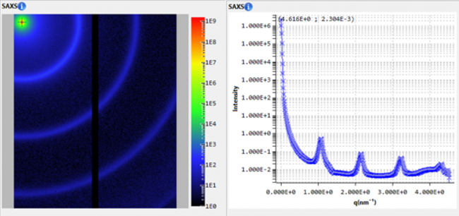
为了更好地解读输出数据,科学家必须拥有直观、可交流和可比较大量图示的工具。
通过将数据处理与高级直观功能的结合,XSACT软件的使用可加快从数据采集到发表的工作流程。
通过内置的移液机械臂和自动软件组件,BioXolver的标准孔盘可以自动装样和测试,为用户带来更高的测试效益。
BioXolver具有强大的数据处理软件BioXTAS RAW,且广泛应用于BioSAXS用户群体。
它可进行基本的数据操作和数据分析(Rg、分子量等),以及使用ATSAS插件的高级分析。
对于高通量分析,自动分析功能可以帮您分析和汇总实验结果。
操作简单。只需将您的样品放置在样品仓里即可。
仪器可进行自动校准,无需用户干预。
自动快捷的数据采集流程。X射线散射数据自动归一化,无需用户校准。这是通过一个嵌入到全自动设备中的强大软件套件,以及独特的固定双探测器配置来实现的。
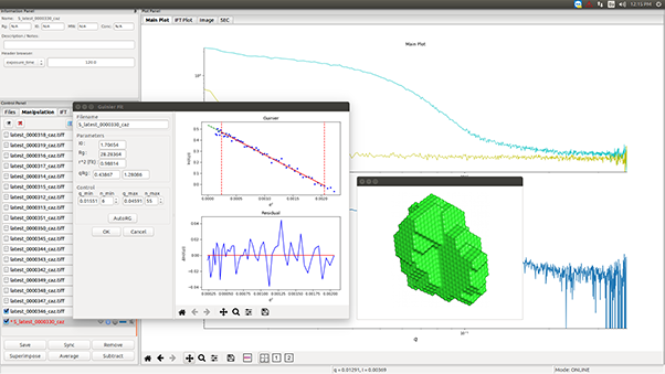
仪器实时显示的精确散射数据可用于进行实时快速的样品反馈,或使用我们的XSACT软件进行进一步的辅助分析。分析功能选择广泛,只需点击几次就能快速获得纳米结构参数。
XSACT生成的高质量可发表的图表,可以通过拖放或保存文件轻松地导出到其他文档。
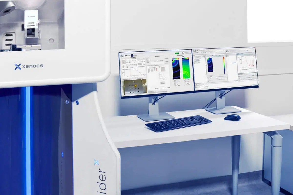
了解更多关于Nano-inXider软件信息,有助于您更快获取样品信息。
Nano-inXider能更快获取结果更简单地进行数据分析
简单易用。智能化的设计可以使研究小角散射的学者,研究材料科学的科学家及技术人员快速掌握。
具有完全的远程操作能力和自动校准,Nano-inXider将人为误差降到最低,并保证重现性和测量可追溯性。
是开放操作实验室的理想选择。
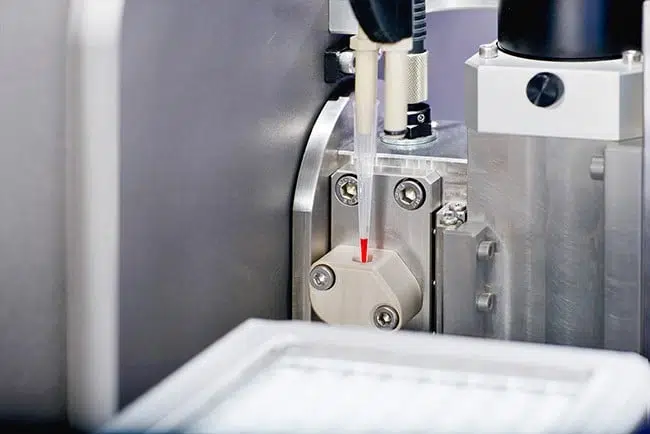
High precision positioning of small sample volumes is ensured by the conjunction of an in-line pipetting robot and the BioCUBE, an advanced capillary flow cell, coupled to machine vision for sample monitoring.
Such low total consumption per sample condition or concentration is highly beneficial for biostructural research.
Tubeless handling of sample volume down to 5 μl. This is unique on the market.
Each sample is first automatically injected into the BioCUBE measurement cell with the tubeless pipetting robot, then positioned into the beam for measurement with dynamic real-time continuous control of its position.
The sample is maintained at the selected temperature both in the well plate during storage and in the BioCUBE during measurements.
Optionally, the BioCUBE may also be provided with integrated UV/Vis capabilities.
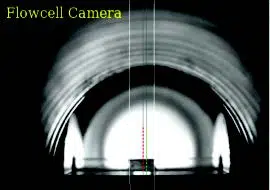
Using machine vision technology, the BioXolver is able to keep a digital eye on the sample at all times.
Once loaded, the sample is positioned with high precision in the X-ray beam while continuous feedback is provided to the automated control software to keep the volume in place.
Simple. Just put your sample in the chamber.
The instrument auto-aligns without any need of user interaction.
Data acquisition workflow is automatic and fast. X-ray scattering data is automatically normalized with no need of calibration by the user. This is achieved through a powerful software suite embedded in a fully motorized equipment using a unique fixed dual detector configuration.
Accurate scattering data instantly displayed by the instrument can be used on the fly for quick sample feedback, or for further assisted analysis using our XSACT software. A large choice of analysis functions is available and quick nanostructure parameters are provided through few clicks.
XSACT produces high quality publication-ready graphs and figures which can be easily exported to other documents through drag-and-drop or saved as files.
Learn more below about the Nano-inXider softwares which is central to obtain quick results on your sample.
The Nano-inXider provides fast answers and straightforward analysis.
It is easy to use. Its SMART design ensures a fast learning curve for both scattering experts, material scientists or technicians.
With full remote operation capability and auto alignment, the Nano-inXider reduces human errors to the minimum, and guarantees reproducibility as well as measurement traceability.
It is ideal for open access labs.
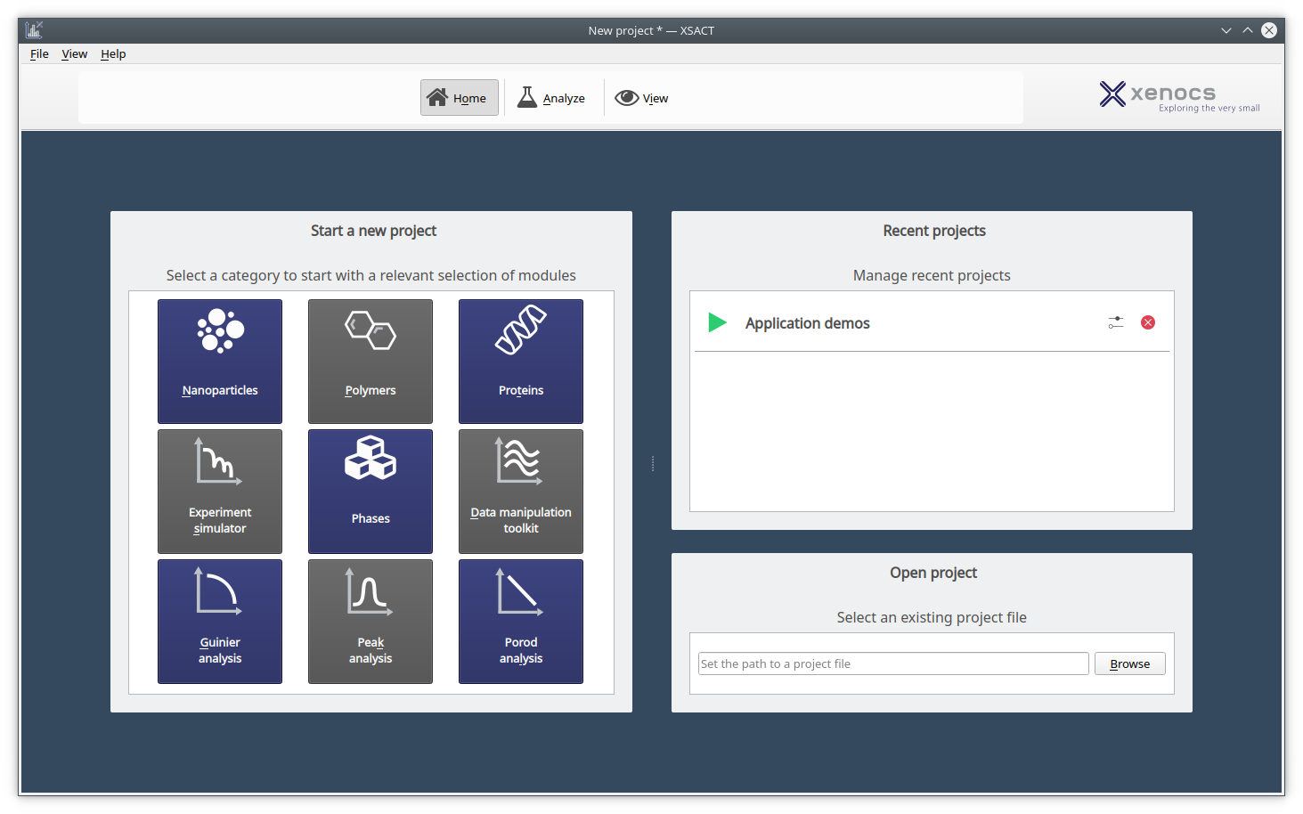
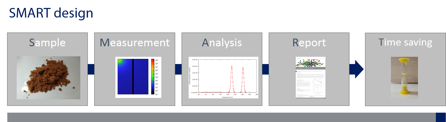
XSACT follows our SMART design for an easy workflow that guides you to results thanks to powerful graphical interactions.
